Power Line Common Fibular Division of the Sciatic Nerve
Human anatomy is not an exact science. For years, grave robbers from multiple countries stole cadavers for dissection in a rush to stake claim on newly discovered muscles.
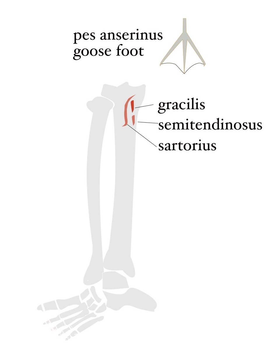 A degree in linguistics would be very helpful in understanding the various independent revelations such as the latin etymology for pes anserinus. The tendons of sartorius, gracilis and semitendinosus muscles attach to the upper medial tibia and resemble the foot of goose. These randomly named anatomical terms serve more to advance the rote memorization of disorganized facts – to pass a test – rather than to understand the relative function of movement.
Even worse, nerves are named like the back roads of Ireland where each county picks the local route name and unit of measure: statute miles (5280ft), kilometers (3280.84ft) or Irish miles (6720ft). The road signs are quite puzzling for a foreigner driving a left-handed stick shift on the ‘wrong’ side of the road trying to navigate a narrow path between a cliff and the mountain rocks blocked by a herd of sheep. Yes, that describes the study of the neurological system in western medicine. Meridians in Chinese Medicine have their own idiosyncrasies.
So you can see that researching human anatomy is like translating at the United Nations, which I always wanted to do when I studied Spanish, Japanese and one semester of German in college. Greek and Latin would have been helpful for word beginnings and endings of human anatomy. Who knew?
The historical annotation of human anatomy was done predominantly by examining cadavers in dissection. Copses don’t move of their own accord, and the muscle attachment sites and nerve lines may have had scars from injuries creating discrepancies in the data. In addition, the lack of movement could not discern wear and tear patterns in the fascia as it was stripped away to better identify the muscles themselves.
Nerves Must Get to Work
A motor neuron signal from the brain causes a muscle to contract or shorten. One end of the muscle serves as an anchor, and the other end of the muscle tendon – attached to the bone near the target joint – pulls the bone in the requested direction. Motor neurons that exit the brain are bundled independent cables that travel through the spinal column. A single cable or motor nerve division is responsible for innervating specific muscles that do specific actions. There may be some overlapping of function within the larger plexus group for emergencies, but mostly, a defined nerve section travels along until it reaches the group of muscles that it administers. A nerve impingement within this scenario will often respond with pain related to a specific motion.
In injury work, there is another very important element of nerves that is often overlooked. After exiting their respective vertebrae, nerves can still have a long way to go to get to work, for instance, a long nerve line assigned to the toes 2-5. For efficiency, the nerves loosely attach to muscles that are heading in the same direction as the destination of the hitch hiker nerve. The nerve may or may not actually innervate the muscles it uses to get to work. If a muscle gets into an accident, such as a hamstring tear, the loosely attached nerve trying to get to work may also get in trouble. However, it will not likely follow the same test patterns used in standard neurological testing. That type of compressed nerve will more often present as a trigger point or an issue somewhere else in the body. A lot more investigative tracking is involved in these very puzzling cases.
After 20 years as a clinical massage therapist, the puzzles get easier to solve. The client history intakes begin to cluster. Oh, you ride horses. You played high school football. One client actually outlined a known pain pattern on the intake form.
Now, it’s time to decrypt the tower of babel and add some intelligence regarding the function of anatomy.
Sacral Plexus
The very long sacral plexus network of motor and sensory neurons exits the spinal column thru vertebrae in the lower back and sacrum, L4-S4. The sciatic nerve is a major part of that bundle with two major divisions, the tibial and common fibular divisions, that innervate the posterior hip and thigh and all of the lower leg and foot. The gluteal muscles are, by definition, innervated by smaller nerve divisions in the sacral plexus, but to say that they are not a broader part of the sciatic nerve within the sacral plexus is merely semantics. To say “I have sciatica.” can really be quite involved.
Common Fibular Division of the Sciatic Nerve
Let’s use anatomy data to evaluate a fairly common musculoskeletal problem faced by aging adults: getting out of chair. If you ask the internet which muscles are used to get out of a chair, the answer is often ‘all of them’. ‘Just try harder’ to strengthen your muscles. The tibial line of the sciatic nerve works with posture muscles that interconnect with gravity allowing humans to be upright. The common fibular line of the sciatic nerve works to move the body upward against gravity such as pushing up out of a squat or getting out of a chair. Muscles related to the common fibular nerve line tend to leverage skeletal strength for power versus muscular strength, such as the hamstrings from the tibial line used for speed.
Conscious Uncoupling of Muscle Groups
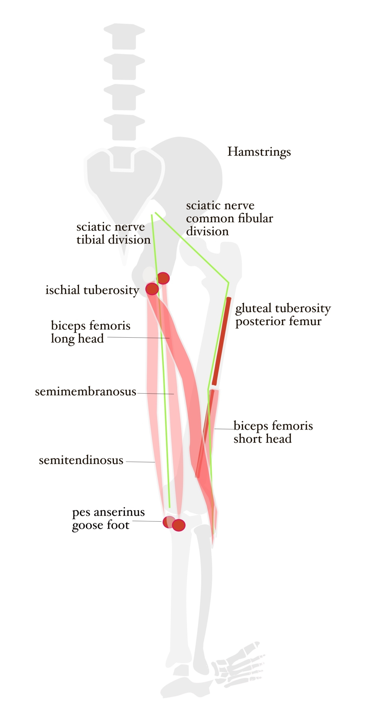
Three out of four hamstring segments are innervated by the tibial nerve division of the sciatic nerve and have long muscles with attachment sites to the bottom of the hip and lower leg bones with no physical attachment to the femur. The short head of the biceps femoris, on the other hand, has a long attachment to the femur bone and is innervated by the fibular nerve division of the sciatic nerve. The short head of the biceps femoris muscle segment is misplaced in the hamstring group even though it shares an attachment site with the long head of the biceps femoris at the top of fibula.
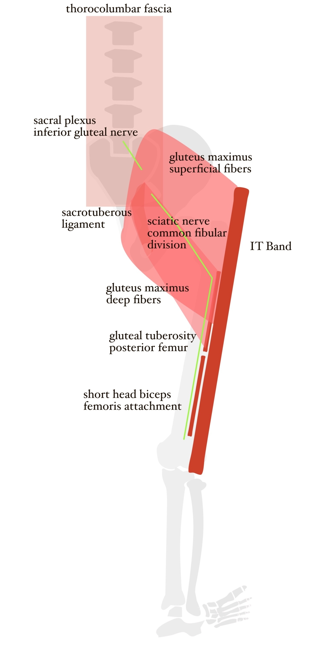
The deep fibers of the gluteus maximus attach to the gluteal tuberosity of the posterior femur directly in line with the short head of the biceps femoris. The superficial fibers of the gluteus maximus are used for hip extension in walking and are innervated by the inferior gluteal nerve, a nerve division of the sacral plexus. The superficial fibers of the gluteus maximus attach to the IT band which offers enhanced range of motion. The deep fibers of the gluteus maximus are much more indicated as a strength position moving against gravity in the secure skeletal attachment. There is also a strong the likelihood that it is innervated by the common fibular nerve division. The deep fibers of the gluteus maximus should be separated as a different muscle from the superficial fibers.
One validation of this theory came from a post polio client. Overall, his body had managed to rid itself of the effects of the polio at age 5 except for a group of muscles in the anterior and lateral compartments of the left lower leg. An investigation along the power line showed that every single muscle attachment section along the entire line of the common fibular nerve division, including the short head of the biceps and the gluteus maximus deep fiber attachment to the femur, were restricted by a heavy concentration of calcium in the tendons. The entire common fibular nerve division of the sciatic nerve was impacted by the polio virus. Why just that nerve line? Does this serve as proof that a deep section of the gluteus maximus as we know it is actually innervated by common fibular nerve division?
Lower Leg Path of Common Fibular Division
The common fibular line passes from the sacrum to the back of the thigh, over the head of the fibula to the lateral and anterior compartments of the lower leg. The tibialis anterior is used in for lifting the foot, anchoring the front leg in a tennis serve or for initiating a roll into an aikido fall. Fibularis longus and brevis make up the lateral compartment of the lower leg and cross the bottom of foot from outside to inside like the stirrup of a horse’s saddle.
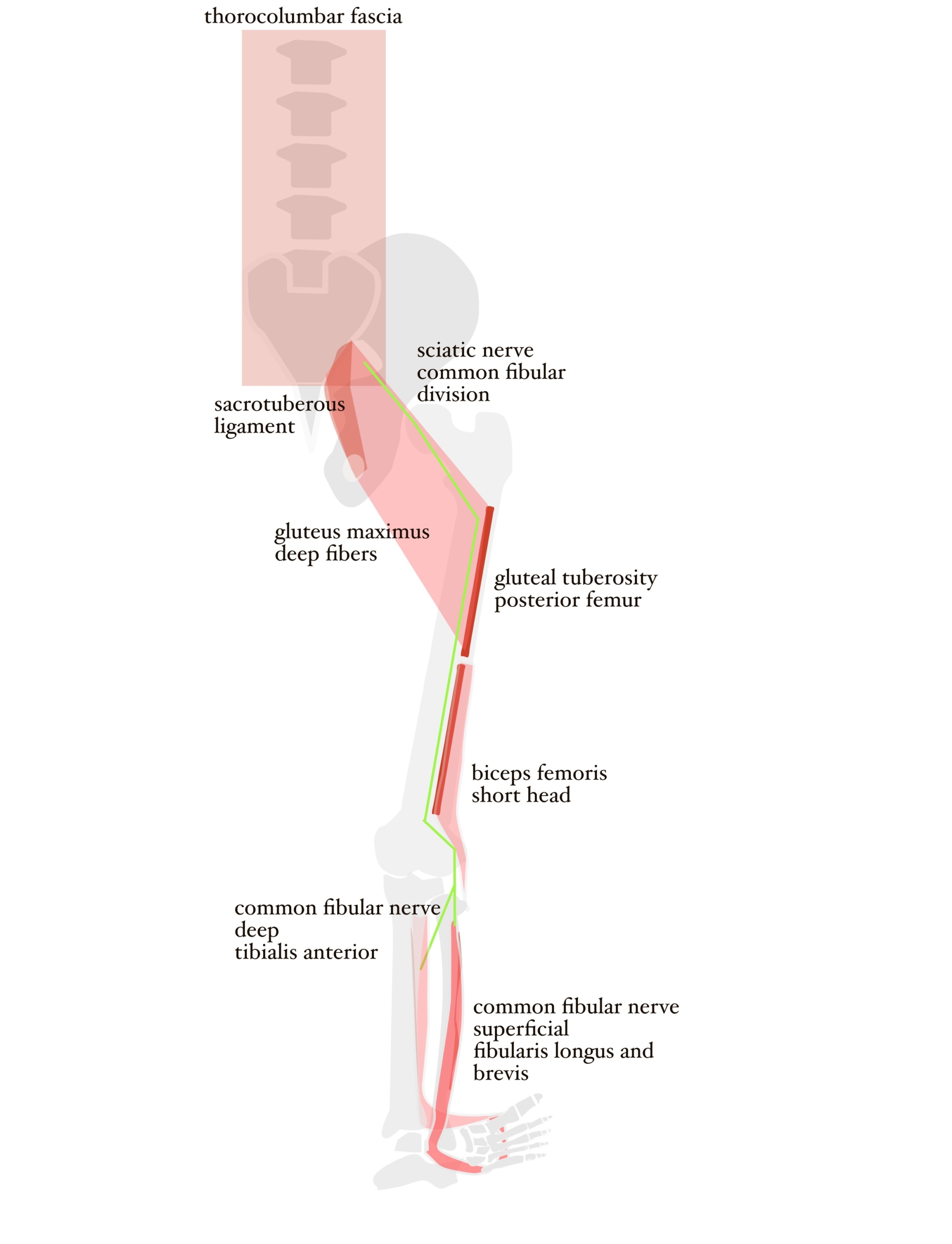
To lift out of a chair, the flat foot is on the ground, and the muscles of the lateral and anterior compartments of the lower leg initiate the upward movement. Next, the short head of the bicep on the common fibular line connects to the deep fibers of the gluteus maximus which connects to the sacrotuberous ligament activating the movement of the entire spinal column through the thorocolumbar fascia. This is a power line to lift against gravity using muscles innervated by the common fibular line.
If a client presents with an inability to get out of a chair, work the muscles along the common fibular nerve line. The long muscle attachment sites along the posterior femur, tibia and fibula can be manipulated using myofascial release, such as foam rolling, or cross fiber friction.
Human anatomy in its flat file data presentation does serve a purpose in education as a discipline of rote memorization to weed out potential practitioners that can’t memorize long, complicated lists. Grouping anatomy and clinical massage session data into intelligent and usable categories can speed the evaluation process of chronic injury to get closer to possible resolution. Clients say my work is magic. It’s really just a long term quest for answers in a jumbled mess of pretentious terminology. It’s easy to make something difficult sound difficult. It’s difficult to make something difficult sound easy. I opt for the latter.
Aut![]() hor: Mary Bai of Tibialis, LLC is practicing Clinical, Orthopedic, Medical Massage in Redwood City, CA.
hor: Mary Bai of Tibialis, LLC is practicing Clinical, Orthopedic, Medical Massage in Redwood City, CA.
Contact: mary@tibialis.com